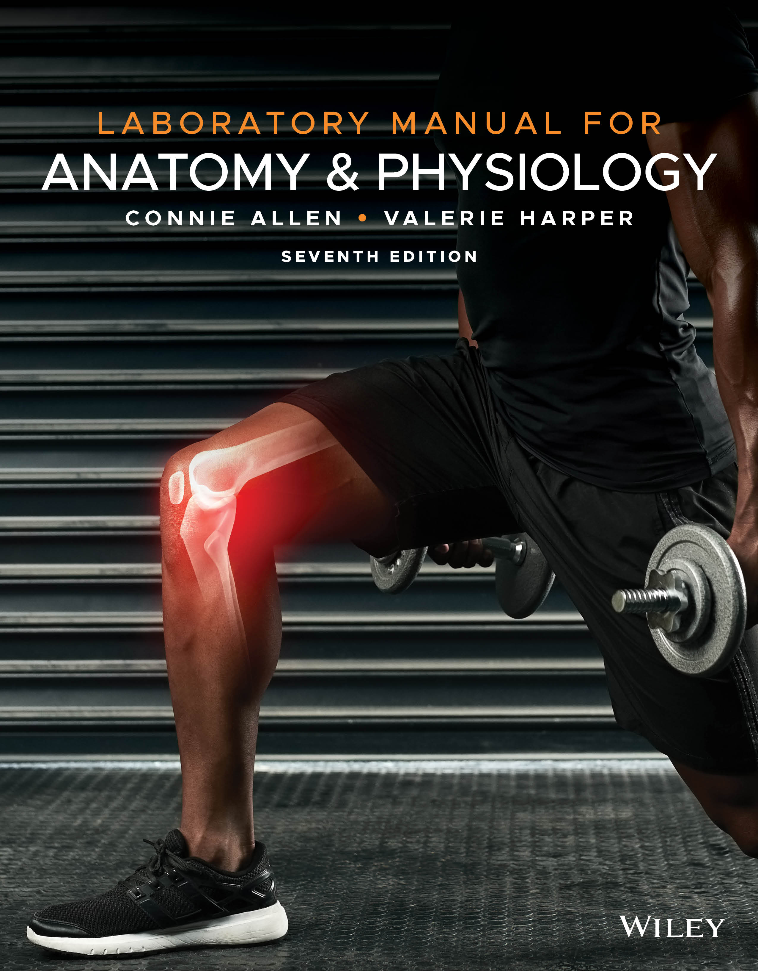
Laboratory Manual for Anatomy & Physiology, 7th Edition
By Connie Allen and Valerie Harper
Laboratory Manual for Anatomy & Physiology, 7th Edition, contains dynamic and applied activities and experiments that help students both visualize anatomical structures and understand complex physiological topics. With many different format options available, and powerful digital resources in WileyPLUS, it’s easy to customize this laboratory manual to best fit your course.
Lab exercises are designed in a way that requires students to first apply information they learned and then critically evaluate it. While the Laboratory Manual for Anatomy and Physiology is designed to complement the latest 16th edition of Principles of Anatomy & Physiology, it can be used with any two-semester A&P text.
Want to learn more about WileyPLUS? Click Here
Create active learners and put students in the driver’s seat with 3D interactives
Assignable with paired assessment questions and fully accessible with interactive keyboard short cuts, 3D Interactives provide an immersive and visually stunning experience that addresses multiple learning modalities. Students can manipulate structures, interact with images, and listen to narrated explanations of physiological concepts to dive deeper into complex physiologic processes.
Help students develop a growth mindset and picture their future career path
Practitioner’s Perspective videos share the perspective of the clinician on a clinical case study. The clinicians represent an array of possible future careers for the students, including doctors and nurses but also EMT, audiologist, acupuncturist, and radiologic technician. The practitioners are diverse and represent broad student populations. Each practitioner shares an engaging story about a person experiencing a disease state such as kidney stones, multiple sclerosis, burns, or a cochlear implant.
Replicate a true-to-life cadaver experience with world-class dissection software
Real Anatomy 2.0 is 3-D imaging software that allows students to dissect through over 40 layers of a real human body to study the anatomical structures of all body systems. Using Real Anatomy, students can interact and practice with a real cadaver as much as they need. Additional dissection videos are also included: in this resource are cat, pig, rat, and human dissection videos. Both the cat and the pig videos include manuals that are available within the resource for download as PDF.
Flip your classroom or recreate the benefits in-person lecture
Created by author Mark Nielsen, Concept Lectures provide a great resource for professors wanting to incorporate more of a flipped classroom approach into their teaching. These dynamic video lectures help students focus on key concepts and make connections to anatomy.
Reinforce techniques used in the laboratory with physiological simulations
PowerPhys 3.0 simulation software allows users to explore physiology principles through 14 self-contained activities. Each activity includes objectives with illustrated and animated review material, pre-lab quizzes, pre-lab reports, data collection and analysis, and a full lab report with discussion and application questions. Experiments contain randomly generated data, allowing users to experiment multiple times but still arrive at the same conclusions.
Bring physiology to life with 3D animations
3-D Physiology uses state-of-the-art 3D visualization to bring the 15 most complicated topics for students to learn and teachers to teach to life. The most requested topic, The Countercurrent Mechanism, helps students visualize a dense, very complex physiological process. Each animation is fully accessible and includes transcript and speed controls allowing students to absorb the material on their own terms.
What’s New:
- Create active learners and put students in the driver’s seat with 3D interactives
Assignable with paired assessment questions and fully accessible with interactive keyboard short cuts, 3D Interactives provide an immersive and visually stunning experience that addresses multiple learning modalities. Students can manipulate structures, interact with images, and listen to narrated explanations of physiological concepts to dive deeper into complex physiologic processes. - Help students develop a growth mindset and picture their future career path
Practitioner’s Perspective videos share the perspective of the clinician on a clinical case study. The clinicians represent an array of possible future careers for the students, including doctors and nurses but also EMT, audiologist, acupuncturist, and radiologic technician. The practitioners are diverse and represent broad student populations. Each of practitioner shares an engaging story about a person who is experiencing a disease state such as kidney stones, multiple sclerosis, burns, or a cochlear implant.

Connie Allen was a Professor of Biology at the Fort Myers Campus of Edison State College for 23 years. She taught human anatomy and physiology and was the coordinator of anatomy and physiology for 20 years. She has been a member of the Human Anatomy and Physiology Society (HAPS) for 25 years and still attends the HAPS annual convention. Connie received her Bachelor’s Degree in Biology from Anderson College and her MS in Anatomy and Physiology from Indiana University Medical Center (IUMC) Indianapolis, Indiana. While at IUMC she also did research using an electron microscope. After graduation, Connie worked for Eli Lilly Co. in Indianapolis, Indiana doing research using the electron microscope and worked in the medical research area conducting clinical trials for an antibiotic that was approved by the FDA.
Initially, Connie planned to continue working at Eli Lilly doing research but discovered that she enjoyed teaching people about her favorite subject: anatomy and physiology. At Edison State College, Connie was the coordinator of the Honors Scholar Program for 5 years and received the Edison President’s Award for Outstanding Professor for Exemplary Service to Students. Her inspiration came from her students — she was dedicated to her students and enjoyed the rewards of teaching at a college with student diversity in academic ability, ethnicity, and age. Connie and her husband love traveling, scuba diving, entertaining people in their home, and enjoying their three children and six grandchildren.

Valerie Harper is a lecturer at Colorado Mesa University at the branch campus in Montrose, CO, where she teaches Anatomy & Physiology and Pathophysiology. She is a member of the Human Anatomy and Physiology Society (HAPS). Valerie received a Bachelor’s Degree in Biology from the University of Miami and a PhD in Physiology from the University of Virginia. At the University of Virginia, Valerie’s research focused on relative amounts of smooth muscle contractile proteins in different tissues and the changes in these proteins that occurred when smooth muscle cells were cultured. She also completed a postdoctoral fellowship at the University of Virginia where she studied the renin-angiotensin system. Valerie pursued a career in research but soon discovered that her real passion is teaching. She started teaching at Edison Community College (now Florida Southwestern State College) where she met Connie Allen. At ECC, she was involved in curriculum development, participated in the Leadership Diversity Program, was nominated for the Thomas Edison Award for Excellence in Teaching, and was recognized in a graduate survey by students as having the most significant influence on them during their enrollment at Edison Community College.
When she is not teaching or editing the Laboratory Manual, Valerie enjoys skiing and paddle boarding, cooking with her husband, as well as attending an occasional Steeler football game with her son or a Penguin hockey game with her daughter.
Exercise 1: Anatomical Language
Exercise 2: Organ Systems and Body Cavities
Exercise 3: Compound Light Microscope
Exercise 4: Cell Structure and Function
Exercise 5: Transport Across the Plasma Membrane
Exercise 6: Tissues
Exercise 7: Integumentary System Structure and Function
Exercise 8: Bone Structure and Function
Exercise 9: Axial Skeleton
Exercise 10: Appendicular Skeleton
Exercise 11: Joints and Synovial Joint Movements
Exercise 12: Skeletal Muscle Structure
Exercise 13: Contraction of Skeletal Muscle
Exercise 14: Skeletal Muscles and Their Actions
Exercise 15: Surface Anatomy
Exercise 16: Nervous Tissue
Exercise 17: Spinal Cord Structure and Function
Exercise 18: Spinal Nerves
Exercise 19: Somatic Reflexes
Exercise 20: Brain Structure and Function
Exercise 21: Cranial Nerves
Exercise 22: Autonomic Nervous System Structure and Function
Exercise 23: General Senses
Exercise 24: Special Senses
Exercise 25: Endocrine Structure and Function
Exercise 26: Blood Components and Blood Tests
Exercise 27: Heart Structure and Function
Exercise 28: Cardiac Cycle
Exercise 29: Blood Vessel Structure and Function
Exercise 30: Blood Vessel Identification
Exercise 31: Lymphatic System Structure and Immune System Function
Exercise 32: Respiratory System Structure and Function
Exercise 33: Pulmonary Ventilation
Exercise 34: Digestive System Structure and Function
Exercise 35: Chemical Digestion
Exercise 36: Urinary System Structure and Function
Exercise 37: Urine Formation and Urinalysis
Exercise 38: Male Reproductive System Structure and Function
Exercise 39: Female Reproductive System Structure and Function
Exercise 40: Human Development
Exercise 41: Heredity

